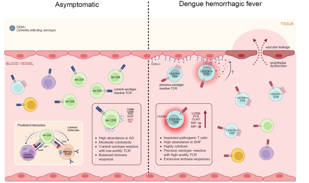
Dengue infects an estimated 390 million people a year, yet the same virus can leave one person asymptomatic and put another in the hospital with dengue hemorrhagic fever (DHF). Antibodies have long taken the blame for that gap, but the role of CD8⁺ “killer” T cells, the immune cells responsible for eliminating virus-infected cells, has stayed unclear. Most studies have examined blood after the acute illness had resolved, when the immune response that shaped disease outcome had already passed.
A team led by co-first authors Sirawit Srikor and Waradon Sungnak, together with Ponpan Matangkasombut, Varodom Charoensawan, and Sarah A. Teichmann went looking during the infection itself. Using HLA tetramers to isolate CD8⁺ T cells targeting a dominant dengue NS3 epitope, combined with plate-based single-cell transcriptomics and paired T cell receptor (TCR) sequencing, the researchers generated one of the most detailed maps to date of antigen-specific CD8⁺ T-cell responses across the clinical spectrum of dengue, from asymptomatic infection to dengue fever (DF) to DHF. In total, they profiled 2,397 virus-specific CD8⁺ T cells, sampled during acute illness and again at convalescence.
The cells turned out to be far from uniform. Five distinct CD8⁺ T-cell states emerged, ranging from resting to highly cytotoxic effector cells. Among them, two subsets were particularly associated with different clinical outcomes. An intermediate (Int) CD8 subset with moderate cytotoxicity was significantly enriched in asymptomatic infection and showed a tendency to recognize peptides from the currently infecting serotype. In contrast, a CX3CR1⁺ CD8⁺ T-cell subset displayed some of the highest TCR binding of any subset, along with elevated expression of cytotoxic genes and a tendency to recognize peptides from serologically inferred previously infecting serotypes. This subset was significantly more abundant in patients with DHF. To place these findings within the broader immune response, the researchers integrated the antigen-specific T-cell data with matched whole-blood single-cell datasets and plasma cytokine profiles. Clonally expanded CX3CR1⁺ CD8⁺ T cells correlated with inflammatory cytokines previously associated with severe dengue, including IL-15 and MIP-1α, consistent with a possible role in vascular leakage, although the study does not establish causation. TCR repertoire analysis further revealed distinct receptor usage between asymptomatic and symptomatic infections, while longitudinal sampling showed CX3CR1⁺ cells predominating during acute infection before Int CD8⁺ cells became more prominent during recovery.
Conducted by researchers at Mahidol University in collaboration with the University of Cambridge, the Wellcome Sanger Institute, the University of Oxford, and the DENFREE Thailand consortium, the study suggests that dengue severity may depend less on the overall magnitude of the CD8⁺ T-cell response than on which functional T-cell subsets dominate during infection. Although the study was limited to selected HLA types, DENV NS3 epitopes, and a relatively modest cohort, it provides important insight into how qualitatively different antigen-specific CD8⁺ T-cell responses may contribute to either protective immunity or immunopathology. These findings provide a framework for future studies and may help guide vaccines and immunotherapies toward protective cellular immunity while minimizing immunopathology.
Explore the Full Study: Nature Communications (2026). https://doi.org/10.1038/s41467-026-73491-5
First Author: Sirawit Srikor and Waradon Sungnak
Co-author: VimvaraVacharathit
Corresponding Author: Ponpan Matangkasombut, Varodom Charoensawan and Sarah A. Teichmann